 Overview A heel spur also known as a calcaneal spur, is a pointed bony outgrowth of the heel bone (calcaneus). Heel spurs do not always cause pain and often are discovered incidentally on X-rays taken for other problems. Heel spurs can occur at the back of the heel and also under the heel bone on the sole of the foot, where they may be associated with the painful foot condition plantar fasciitis. Causes Athletes who participate in sports that involve a significant amount of jumping and running on hard surfaces are most likely to suffer from heel spurs. Some other risk factors include poor form while walking which can lead to undue stress on the heel and its nerves and ligaments. Shoes that are not properly fitted for the wearer?s feet. Poor arch support in footwear. Being overweight. Occupations that require a lot of standing or walking. Reduced flexibility and the thinning of the fat pad along the bottom of the heel, both of which are a typical depreciation that comes with aging.  Symptoms Bone spurs may cause sudden, severe pain when putting weight on the affected foot. Individuals may try to walk on their toes or ball of the foot to avoid painful pressure on the heel spur. This compensation during walking or running can cause additional problems in the ankle, knee, hip, or back. Diagnosis Diagnosis is made using a few different technologies. X-rays are often used first to ensure there is no fracture or tumor in the region. Then ultrasound is used to check the fascia itself to make sure there is no tear and check the level of scar tissue and damage. Neurosensory testing, a non-painful nerve test, can be used to make sure there is not a local nerve problem if the pain is thought to be nerve related. It is important to remember that one can have a very large heel spur and no plantar fasciitis issues or pain at all, or one can have a great deal of pain and virtually no spur at all. Non Surgical Treatment FIRST, Reduce the acute pain. This is done by a combination of several things; injection of a synthetic relative of cortisone into the heel, a prescription of anti-inflammatory pills to reduce inflammation, physical therapy and a special heel pad. About 50% of the time, these treatments will permanently relieve the pain. In the other 50%, the pain becomes recurrent, and the treatment proceeds to Stage II. SECOND, Recurrent, painful heel spur is caused by the tug and pull of the plantar fascia ligament on the heel bone with each step. When the pain is recurrent, arch supports are made to prevent sagging of the arch. The arch supports are custom-made according to the size and shape of the feet. This prevents the arch from sagging and the ligament from tugging and pulling on the heel bone. The inflammation and pain eventually go away as the first phase of treatment is continued along with the arch supports, although the spur itself remains. THIRD, Surgery to remove the spur is possible and is usually done as Day Surgery. Surgical Treatment When chronic heel pain fails to respond to conservative treatment, surgical treatment may be necessary. Heel surgery can provide relief of pain and restore mobility. The type of procedure used is based on examination and usually consists of releasing the excessive tightness of the plantar fascia, called a plantar fascia release. Depending on the presence of excess bony build up, the procedure may or may not include removal of heel spurs. Similar to other surgical interventions, there are various modifications and surgical enhancements regarding surgery of the heel. Prevention Heel spurs and plantar fasciitis can only be prevented by treating any underlying associated inflammatory disease. Overview
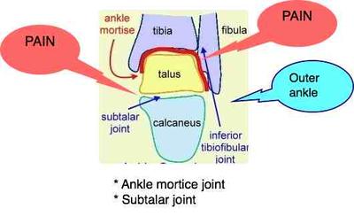
Bursas are small fluid containing sacs, that are situated between areas of high friction such as bone against the floor (heel) and bone against other soft tissue structures like tendons, skin and or muscle. The bursa job is to act as a shock absorber, and to allow stress free movement between the above noted structures. Bursitis is a swellinginflammation of the bursa sac, due to constant micro trauma or overuse. In the foot Abnormal Pronation, most often caused by Morton?s Toe. Causes Posterior heel pain can come from one of several causes. When a physician is talking about posterior heel pain, he or she is referring to pain behind the heel, not below it. Pain underneath the heel, on the bottom of the foot, has several causes including Tarsal Tunnel Syndrome. Plantar Fasciitis. Heel Spurs. Symptoms Patients with this condition typically experience pain at the back of the ankle and heel where the Achilles tendon attaches into the heel bone. Pain is typically experienced during activities requiring strong or repetitive calf contractions (often involving end of range ankle movements) such as walking (especially uphill), going up and down stairs, running, jumping or hopping (especially whilst wearing excessively tight shoes). Often pain may be worse with rest after these activities (especially that night or the following morning). The pain associated with this condition may 'warm up' with activity in the initial stages of injury. As the condition progresses, patients may experience symptoms that increase during sport or activity, affecting performance. In severe cases, patients may walk with a limp or be unable to weight bear on the affected leg. Other symptoms may include tenderness on firmly touching the affected bursa and swelling around the Achilles region. Diagnosis The doctor will discuss your symptoms and visually assess the bones and soft tissue in your foot. If a soft tissue injury is suspected, an MRI will likely be done to view where and how much the damage is in your ankle. An x-ray may be recommended to rule out a bone spur or other foreign body as the cause of your ankle pain. As the subcutaneous bursa is close to the surface of the skin, it is more susceptible to septic, or infectious, bursitis caused by a cut or scrape at the back of the heel. Septic bursitis required antibiotics to get rid of the infection. Your doctor will be able to determine whether there is an infection or not by drawing a small sample of the bursa fluid with a needle. Non Surgical Treatment Many cases of retrocalcaneal bursitis can be resolved with self-care that is focused on reducing inflammation and eliminating activities or positions that aggravate the bursa. Some cases, however, may become more serious and require more medical interventions. Rarely, surgery is needed. Following the R.I.C.E. formula, or Rest, Ice, Compression, and Elevation, is often sufficient to treat aseptic bursitis. Rest. People with retrocalcaneal bursitis should avoid activities that irritate the bursa, such as jogging or excessive walking. Ice. Applying a cold compress to the back of the ankle for about 20 minutes two or three times a day may help alleviate symptoms and decrease swelling. Compression. An elastic medical bandage (e.g. Ace? bandage) wrapped around the affected heel and ankle can help control swelling. Elevating the affected heel. Sitting down with the leg elevated on a stool or lying down with the foot elevated on a pillow can help reduce blood flow to the area, thereby reducing inflammation. Surgical Treatment Surgery is rarely need to treat most of these conditions. A patient with a soft tissue rheumatic syndrome may need surgery, however, if problems persist and other treatment methods do not help symptoms. Prevention You can avoid the situation all together if you stop activity as soon as you see, and feel, the signs. Many runners attempt to push through pain, but ignoring symptoms only leads to more problems. It?s better to take some time off right away than to end up taking far more time off later. Runners aren?t the only ones at risk. The condition can happen to any type of athlete of any age. For all you women out there who love to wear high-heels-you?re at a greater risk as well. Plus, anyone whose shoes are too tight can end up with calcaneal bursitis, so make sure your footwear fits. If the outside of your heel and ankle hurts, calcaneal bursitis could be to blame. Get it checked out.  Overview OverviewHammer toe is often a harmless and painless condition. Although the toe may be curled permanently, hammertoe should not cause any long-term problems other than a more difficult time finding shoes that fit. If hammertoe is treated and preventive measures are followed, the condition should not return. Wearing tight or constricting shoes can cause hammertoe to return. Causes Hammer toes result from a muscle imbalance which causes the ligaments and tendons to become unnaturally tight. This results in the joint curling downward. Arthritis can also lead to many different forefoot deformities, including hammer toes.  Symptoms SymptomsHammer toes can cause problems with walking and lead to other foot problems, such as blisters, calluses, and sores. Pain is caused by constant friction over the top of the toe?s main joint. It may be difficult to fit into some shoe gear due to the extra space required for the deformed toe. In many cases there will be pain on the ball of the foot over the metatarsals along with callus formation. This is due to the toes not functioning properly, failing to properly touch the ground during the gait cycle. The ball of hammertoes the foot then takes the brunt of the ground forces, which causes chronic pain. Diagnosis Some questions your doctor may ask of you include, when did you first begin having foot problems? How much pain are your feet or toes causing you? Where is the pain located? What, if anything, seems to improve your symptoms? What, if anything, appears to worsen your symptoms? What kind of shoes do you normally wear? Your doctor can diagnose hammertoe or mallet toe by examining your foot. Your doctor may also order X-rays to further evaluate the bones and joints of your feet and toes. Non Surgical Treatment Your doctor will decide what type of hammertoe you have and rule out other medical conditions. Treatment may range from more appropriate footgear to periodic trimming and padding of the corn. Cortisone injections may be indicated if a bursitis is present. Antibiotics may be utilized in the presence of infection. Removable accommodative pads may be made for you. Surgical Treatment Toe Relocation procedures are ancillary procedures that are performed in conjunction with one of the two methods listed about (joint resection or joint mending). When the toe is deformed (buckled) at the ball of the foot, then this joint often needs to be re-positioned along with ligament releases/repair to get the toe straight. A temporary surgical rod is needed to hold the toe aligned while the ligaments mend.  Overview OverviewHammer toes a bending and hardening of the joints Hammer toe of the second, third, fourth, or fifth toes. If you look down at your feet and you can?t see the tips of the toenails, you might suffer from hammertoe. Early signs of hammertoe are a bend in the joint of any toe except the big toe. The bend in the joint causes the top of the toe to appear to curl under as if it?s ?hammering? into the floor. Causes More often than not, wearing shoes that do not fit a person well for too long may actually cause hammer toes. Wearing shoes that are too narrow or too tight for the person for extended periods of time may eventually take a toll on the person's feet. The same is true for women who like wearing high-heeled shoes with narrow toe boxes.  Symptoms SymptomsThe most obvious symptom of hammertoe is the bent, hammer-like or claw-like appearance of one or more of your toes. Typically, the proximal joint of a toe will be bending upward and the distal joint will be bending downward. In some cases, both joints may bend downward, causing the toes to curl under the foot. In the variation of mallet toe, only the distal joint bends downward. Other symptoms may include Pain and stiffness during movement of the toe, Painful corns on the tops of the toe or toes from rubbing against the top of the shoe's toe box, Painful calluses on the bottoms of the toe or toes, Pain on the bottom of the ball of the foot, Redness and swelling at the joints. If you have any of these symptoms, especially the hammer shape, pain or stiffness in a toe or toes, you should consider consulting your physician. Even if you're not significantly bothered by some of these symptoms, the severity of a hammertoe can become worse over time and should be treated as soon as possible. Up to a point hammertoes can be treated without surgery and should be taken care of before they pass that point. After that, surgery may be the only solution. Diagnosis The earlier a hammertoe is diagnosed, the better the prognosis and treatment options. Your doctor will be able to diagnose your hammertoe with a simple examination of the foot and your footwear. He or she may take an x-ray to check the severity of the condition. You may also be asked about your symptoms, your normal daily activities, and your medical and family history. Non Surgical Treatment Any forefoot problems that cause pain or discomfort should be given prompt attention. Ignoring the symptoms can aggravate the condition and lead to a breakdown of tissue, or possibly even infection. Conservative treatment of mallet toes begins with accommodating the deformity. The goal is to relieve pressure, reduce friction, and transfer forces from the sensitive areas. Shoes with a high and broad toe box (toe area) are recommended for people suffering from forefoot deformities such as mallet toes. This prevents further irritation in the toe area from developing. Other conservative treatment includes forefoot supports such as gel toe caps, gel toe shields and toe crests. Gel forefoot supports provide immediate comfort and relief from common forefoot disorders without drying the skin. Surgical Treatment Surgical correction is necessary in more severe cases and may consist of removing a bone spur (exostectomy) removing the enlarged bone and straightening the toe (arthroplasty), sometimes with internal fixation using a pin to realign the toe; shortening a long metatarsal bone (osteotomy) fusing the toe joint and then straightening the toe (arthrodesis) or simple tendon lengthening and capsule release in milder, flexible hammertoes (tenotomy and capsulotomy). The procedure chosen depends in part on how flexible the hammertoe is.  Prevention PreventionWalking barefoot increases the risk for injury and infection. Being on your feet throughout the day can cause them to swell, this is the best time to buy shoes to get a better fit. Do not buy shoes that feel tight. Do not buy shoes that ride up and down your heel as you walk. The ball of your foot should fit into the widest part of the shoe. Remember, the higher the heel the less safe the shoe will be. Avoid shoes with pointed or narrow toes. If the shoes hurt, do not wear them. If you start noticing the beginning signs of hammer toes, you may still be able to prevent the tendons from tightening by soaking your feet every day in warm water, wearing toe friendly shoes, and performing foot exercises such as stretching your toes and ankles. A simple exercise such as placing a small towel on the floor and then picking it up using only your toes can help to restore the flexibility of tendons.
Overview
 Even though bunions are a common foot deformity, there are misconceptions about them. Many people may unnecessarily suffer the pain of bunions for years before seeking treatment. A bunion (also referred to as hallux valgus or hallux abducto valgus) is often described as a bump on the side of the big toe. But a bunion is more than that. The visible bump actually reflects changes in the bony framework of the front part of the foot. The big toe leans toward the second toe, rather than pointing straight ahead. This throws the bones out of alignment, producing the bunion?s ?bump.? Bunions are a progressive disorder. They begin with a leaning of the big toe, gradually changing the angle of the bones over the years and slowly producing the characteristic bump, which becomes increasingly prominent. Symptoms usually appear at later stages, although some people never have symptoms. Even though bunions are a common foot deformity, there are misconceptions about them. Many people may unnecessarily suffer the pain of bunions for years before seeking treatment. A bunion (also referred to as hallux valgus or hallux abducto valgus) is often described as a bump on the side of the big toe. But a bunion is more than that. The visible bump actually reflects changes in the bony framework of the front part of the foot. The big toe leans toward the second toe, rather than pointing straight ahead. This throws the bones out of alignment, producing the bunion?s ?bump.? Bunions are a progressive disorder. They begin with a leaning of the big toe, gradually changing the angle of the bones over the years and slowly producing the characteristic bump, which becomes increasingly prominent. Symptoms usually appear at later stages, although some people never have symptoms.Causes With prolonged wearing of constraining footwear your toes will adapt to the new position and lead to the deformity we know as a foot bunion. Footwear is not the only cause of a bunion. Injuries to the foot can also be a factor in developing a bunion. Poor foot arch control leading to flat feet or foot overpronation does make you biomechanically susceptible to foot bunions. A family history of bunions also increases your likelihood of developing bunions. Many people who have a bunion have a combination of factors that makes them susceptible to having this condition. For example, if you are a women over the age of forty with a family history of bunions, and often wear high-heeled shoes, you would be considered highly likely to develop a bunion. Symptoms Bunions starts as the big toe begins to deviate, developing a firm bump on the inside edge of the foot, at the base of the big toe. Initially, at this stage the bunion may not be painful. Later as the toes deviate more the bunion can become painful, there may be redness, some swelling, or pain at or near the joint. The pain is most commonly due to two things, it can be from the pressure of the footwear on the bunion or it can be due to an arthritis like pain from the pressure inside the joint. The motion of the joint may be restricted or painful. A hammer toe of the second toe is common with bunions. Corns and calluses can develop on the bunion, the big toe and the second toe due to the alterations in pressure from the footwear. The pressure from the great toe on the other toes can also cause corns to develop on the outside of the little toe or between the toes. The change in pressure on the toe may predispose to an ingrown nail. Diagnosis Your doctor will ask questions about your past health and carefully examine your toe and joint. Some of the questions might be: When did the bunions start? What activities or shoes make your bunions worse? Do any other joints hurt? The doctor will examine your toe and joint and check their range of motion. This is done while you are sitting and while you are standing so that the doctor can see the toe and joint at rest and while bearing weight. X-rays are often used to check for bone problems or to rule out other causes of pain and swelling. Other tests, such as blood tests or arthrocentesis (removal of fluid from a joint for testing), are sometimes done to check for other problems that can cause joint pain and swelling. These problems might include gout , rheumatoid arthritis , or joint infection. Non Surgical Treatment Pain is the main reason that you seek treatment for bunion. Analgesics may help. Inflammation it best eased via ice therapy and techniques or exercises that deload the inflammed structures. Anti-inflammatory medications may help. Your physiotherapist will use an array of treatment tools to reduce your pain and inflammation. These include ice, electrotherapy, acupuncture, deloading taping techniques, soft tissue massage and orthotics to off-load the bunion. As your pain and inflammation settles, your physiotherapist will turn their attention to restoring your normal toe and foot joint range of motion and muscle length. Treatment may include joint mobilisation and alignment techniques, massage, muscle and joint stretches, taping, a bunion splint or orthotic. Your physiotherapist is an expert in the techniques that will work best for you. Your foot posture muscles are vital to correct the biomechanics that causing your bunion to deteriorate. Your physiotherapist will assess your foot posture muscles and prescribe the best exercises for you specific to your needs. During this stage of your rehabilitation is aimed at returning you to your desired activities. Everyone has different demands for their feet that will determine what specific treatment goals you need to achieve. For some it be simply to walk around the block. Others may wish to run a marathon or return to a labour-intensive activity. Your physiotherapist will tailor your rehabilitation to help you achieve your own functional goals. Bunions will deform further with no attention. Plus, the bunion pain associated does have a tendency to return. The main reason is biomechanical. In addition to your muscle control, your physiotherapist will assess your foot biomechanics and may recommend either a temporary off-the shelf orthotic or refer you to a podiatrist for custom made orthotics. You should avoid wearing high heel shoes and shoes with tight or angular toe boxes. Your physiotherapist will guide you.  Surgical Treatment The primary goal of bunion surgery is to relieve the pain associated with the deformity. This is accomplished by correcting the underlying abnormal metatarsal position by realigning it toward the second toe. Removing excessive bone formation on the bunion "bump", releasing the soft tissue tightness which is pulling the big toe towards the second toe. Tightening the soft tissues which are overly stretched on the bump side of the joint. Re-establish the correct alignment of the cartilage surfaces. Move the sesamoid bones into correct alignment. Realign the great toe. Bunion surgery procedures are based on many factors, including health, age and lifestyle of the patient. However, a critical factor in procedure choice is the grading of the bunion deformity. Prevention Bunions often become painful if they are allowed to progress. But not all bunions progress. Many bunion problems can be managed without surgery. In general, bunions that are not painful do not need surgical correction. For this reason, orthopaedic surgeons do not recommend ?preventive? surgery for bunions that do not hurt, with proper preventive care, they may never become a problem. Overview
Normal, healthy feet pronate! Normal pronation does not need to be ?corrected?. However, some people OVER-pronate. Those people need a shoe that supports their over-pronating foot to help guide the foot and avoid injury. So, what does pronation mean exactly? Well, ?pronate? is the word used to describe the natural motion of the foot after it strikes the ground. When a person with a normally pronating foot runs, the outside part of the heel strikes the ground. As the individual shifts the body weight forward, the foot rolls inward (pronates) and the entire foot comes into contact with the ground. This allows the foot to properly support the body and absorb the impact forces. Motion continues forward and the peron pushes off (called ?toe off?) evenly from the front of the foot. Someone who OVER-pronates strikes the ground with the heel in the same way, but the foot rolls too far inward (overpronation). This causes foot and ankle strain, as it does not allow the foot and ankle to properly support the body nor to properly absorb the impact forces. As motion continues forward, they will toe-off more from the ball of her foot. Runners who overpronate are susceptible to foot, ankle and knee problems if they don't wear a shoe that properly supports the motion of their feet.  Causes There are many possible causes for overpronation, but researchers have not yet determined one underlying cause. Hintermann states, Compensatory overpronation may occur for anatomical reasons, such as a tibia vara of 10 degrees or more, forefoot varus, leg length discrepancy, ligamentous laxity, or because of muscular weakness or tightness in the gastrocnemius and soleus muscles. Pronation can be influenced by sources outside of the body as well. Shoes have been shown to significantly influence pronation. Hintermann states that the same person can have different amounts of pronation just by using different running shoes. It is easily possible that the maximal ankle joint eversion movement is 31 degrees for one and 12 degrees for another running shoe. Symptoms Because pronation is a twisting of the foot, all of the muscles and tendons which run from the leg and ankle into the foot will be twisted. In over-pronation, resulting laxity of the soft tissue structures of the foot and loosened joints cause the bones of the feet shift. When this occurs, the muscles which attach to these bones must also shift, or twist, in order to attach to these bones. The strongest and most important muscles that attach to our foot bones come from our lower leg. So, as these muscles course down the leg and across the ankle, they must twist to maintain their proper attachments in the foot. Injuries due to poor biomechanics and twisting of these muscles due to over-pronation include: shin splints, Achilles Tendonitis, generalized tendonitis, fatigue, muscle aches and pains, cramps, ankle sprains, and loss of muscular efficiency (reducing walking and running speed and endurance). Foot problems due to over-pronation include: bunions, heel spurs, plantar fasciitis, fallen and painful arches, hammer toes, and calluses. Diagnosis Firstly, look at your feet in standing, have you got a clear arch on the inside of the foot? If there is not an arch and the innermost part of the sole touches the floor, then your feet are over-pronated. Secondly, look at your running shoes. If they are worn on the inside of the sole in particular, then pronation may be a problem for you. Thirdly, try the wet foot test. Wet your feet and walk along a section of paving and look at the footprints you leave. A normal foot will leave a print of the heel, connected to the forefoot by a strip approximately half the width of the foot on the outside of the sole. If you?re feet are pronated there may be little distinction between the rear and forefoot, shown opposite. The best way to determine if you over pronate is to visit a podiatrist or similar who can do a full gait analysis on a treadmill or using forceplates measuring exactly the forces and angles of the foot whilst running. It is not only the amount of over pronation which is important but the timing of it during the gait cycle as well that needs to be assessed. Non Surgical Treatment An orthotic is a device inserted inside the shoe to assist in prevention and/or rehabilitation of injury. Orthotics support the arch, prevent or correct functional deformities, and improve biomechanics. Prescription foot orthoses are foot orthoses which are fabricated utilizing a three dimensional representation of the plantar foot and are specifically constructed for an individual using both weightbearing and nonweightbearing measurement parameters and using the observation of the foot and lower extremity functioning during weightbearing activities. Non-prescription foot orthoses are foot which are fabricated in average sizes and shapes in an attempt to match the most prevalent sizes and shapes of feet within the population without utilizing a three dimensional representation of the plantar foot of the individual receiving the orthosis. Prevention With every step we take, we place at least half of our body weight on each foot (as we walk faster, or run, we can exert more than twice our body weight on each foot). As this amount of weight is applied to each foot there is a significant shock passed on to our body. Custom-made orthotics will absorb some of this shock, helping to protect our feet, ankles, knees, hips, and lower back. Overview
Calcaneal apophysitis (Sever?s disease) is the most common cause of heel pain in young athletes. Calcaneal apophysitis is a painful inflammation of the heel?s calcaneal apophysis growth plate, believed to be caused by repetitive microtrauma from the pull of the Achilles tendon on the apophysis. Patients with calcaneal apophysitis may have activity-related pain in the posterior aspect of the heel. 60 percent of patients report bilateral pain. This condition is usually treated conservatively with stretching and arch supports. The young athlete should be able to return to normal activities as the pain decreases. Calcaneal apophysitis (Sever?s Disease) may last for months. Increasing pain, despite measures listed below, warrants a return visit to the physician. Causes Physically active children run the risk of developing Sever?s disease because they put the most strain on their growing bones. Sever?s usually occurs during the adolescent growth spurt, when young people grow most rapidly. (This growth spurt can begin any time between the ages of 8 and 13 for girls and 10 and 15 for boys.) By age 15 the back of the heel usually finished growing. As teens grow, the growth plates harden and the growing bones fuse together into mature bone. Young people engaged in physical activities and sports that involve jumping and running on hard surfaces-such as track, basketball, soccer, and gymnastics-are ata higher risk for developing Sever?s disease. Poor-fitting shoes can contribute by not providing enough support or padding for the feet or by rubbing against the back of the heel. Symptoms The typical patient is a child between 10 and 13 years of age, complaining of pain in one or both heels with running and walking. The pain is localized to the point of the heel where the tendo-Achilles inserts into the calcaneus, and is tender to deep pressure at that site. Walking on his toes relieves the pain. Diagnosis A doctor can usually tell that a child has Sever's disease based on the symptoms reported. To confirm the diagnosis, the doctor will probably examine the heels and ask about the child's activity level and participation in sports. The doctor might also use the squeeze test, squeezing the back part of the heel from both sides at the same time to see if doing so causes pain. The doctor might also ask the child to stand on tiptoes to see if that position causes pain. Although imaging tests such as X-rays generally are not that helpful in diagnosing Sever's disease, some doctors order them to rule out other problems, such as fractures. Sever's disease cannot be seen on an X-ray. Non Surgical Treatment Depending on the diagnosis and the severity of the pain, there a number of treatment options available. Rest, reduce activity, your child should reduce or stop any activity that causes pain, such as sports and running. This can be a difficult option, as children are normally quite willful in pursuit of their favorite pastimes. Over the counter anti-inflammatory drugs, such as ibuprofen (found in Nurofen), to help reduce pain and inflammation. Make certain your child does stretching exercises before play. This will often help reduce the stress on the fascia and relieve heel pain in your child. Orthotic insoles. Orthotics made for children will help support the foot properly, and help prevent over-pronation or improper gait by supporting your child?s foot into a proper biomechanical position. Do not hesitate to schedule an appointment with a podiatrist, should your child?s heel and foot pain persist.
Overview
 Achilles tendon rupture is when the achilles tendon breaks. The achilles is the most commonly injured tendon. Rupture can occur while performing actions requiring explosive acceleration, such as pushing off or jumping. The male to female ratio for Achilles tendon rupture varies between 7:1 and 4:1 across various studies. Achilles tendon rupture is when the achilles tendon breaks. The achilles is the most commonly injured tendon. Rupture can occur while performing actions requiring explosive acceleration, such as pushing off or jumping. The male to female ratio for Achilles tendon rupture varies between 7:1 and 4:1 across various studies.Causes Factors that may increase your risk of Achilles tendon rupture include some of the following. Age. The peak age for Achilles tendon rupture is 30 to 40. Sex. Achilles tendon rupture is up to five times more likely to occur in men than in women. Recreational sports. Achilles tendon injuries occur more often during sports that involve running, jumping, and sudden starts and stops, such as soccer, basketball and tennis. Steroid injections. Doctors sometimes inject steroids into an ankle joint to reduce pain and inflammation. However, this medication can weaken nearby tendons and has been associated with Achilles tendon ruptures. Certain antibiotics. Fluoroquinolone antibiotics, such as ciprofloxacin (Cipro) or levofloxacin (Levaquin), increase the risk of Achilles tendon rupture. Symptoms The classic sign of an Achilles' tendon rupture is a short sharp pain in the Achilles' area, which is sometimes accompanied by a snapping sound as the tendon ruptures. The pain usually subsides relatively quickly into an aching sensation. Other signs that are likely to be present subsequent to a rupture are the inability to stand on tiptoe, inability to push the foot off the ground properly resulting in a flat footed walk. With complete tears it may be possible to feel the two ends of tendon where it has snapped, however swelling to the area may mean this is impossible. Diagnosis The diagnosis of an Achilles tendon rupture can be made easily by an orthopedic surgeon. The defect in the tendon is easy to see and to palpate. No x-ray, MRI or other tests are necessary. Non Surgical Treatment Your doctor will advise you exactly when to start your home physical therapy program, what exercises to do, how much, and for how long to continue them. Alphabet Range of Motion exercises. Typically, the first exercise to be started (once out of a non-removable cast). While holding your knee and leg still (or cross your leg), you simply write the letters of the alphabet in an imaginary fashion while moving your foot and ankle (pretend that the tip of your toe is the tip of a pencil). Motion the capital letter A, then B, then C, all the way through Z. Do this exercise three times per day (or as your doctor advises). Freeze a paper cup with water, and then use the ice to massage the tendon area as deeply as tolerated. The massage helps to reduce the residual inflammation and helps to reduce the scarring and bulkiness of the tendon at the injury site. Do the ice massage for 15-20 minutes, three times per day (or as your doctor advises). Calf Strength exercises. This exercise is typically delayed and not used in the initial stages of rehabilitation, begin only when your doctor advises. This exercise is typically done while standing on just the foot of the injured side. Sometimes, the doctor will advise you to start with standing on both feet. Stand on a step with your forefoot on the step and your heel off the step. The heel and forefoot should be level (neither on your tip toes nor with your heel down). Lower your heel very slowly as low as it will go, then rise back up to the level starting position, again very slowly. This is not a fast exercise. Repeat the exercise as tolerated. The number of repetitions may be very limited at first. Progress the number of repetitions as tolerated. Do this exercise one to two times per day (or as your doctor advises).  Surgical Treatment Surgery is recommended to those who are young to middle-aged and active. The ruptured tendon is sewn together during surgery. This is an outpatient procedure. Afterward the leg is put into a splint cast or walking boot. Physical therapy will be recommended. In about 4 to 6 months, healing is nearly complete. However, it can take up to a year to return to sports fully. Prevention To reduce your chance of developing Achilles tendon problems, follow the following tips. Stretch and strengthen calf muscles. Stretch your calf to the point at which you feel a noticeable pull but not pain. Don't bounce during a stretch. Calf-strengthening exercises can also help the muscle and tendon absorb more force and prevent injury. Vary your exercises. Alternate high-impact sports, such as running, with low-impact sports, such as walking, biking or swimming. Avoid activities that place excessive stress on your Achilles tendons, such as hill running and jumping activities. Choose running surfaces carefully. Avoid or limit running on hard or slippery surfaces. Dress properly for cold-weather training and wear well-fitting athletic shoes with proper cushioning in the heels. Increase training intensity slowly. Achilles tendon injuries commonly occur after abruptly increasing training intensity. Increase the distance, duration and frequency of your training by no more than 10 percent each week. |
